

Objectives
Exposure of humans to microgravity affects cells and tissues at a variety of levels. Musculoskeletal changes (e.g. significant bone and muscle loss) occur even when astronauts exercise regularly, but the mechanisms are not yet understood. Previous flight experiments examined cells in monolayers and lasted 6-28 days. Tissue engineering, a new field enabling three-dimensional tissue equivalents to be created from isolated cells in conjunction with biomaterials and bioreactor culture vessels, can provide a basis for systematic, controlled in vitro studies. Cartilage was selected as a model musculoskeletal tissue for this first long-term space study because of its resilience and low metabolic requirements. Our working hypothesis was that in vitro cartilage formation is affected by space flight. Our specific objectives were two fold: (1) to maintain cell viability for four months in space and (2) to study effects of the space environment on the growth and function of tissue engineered cartilage.
Shuttle-Mir Missions Approach
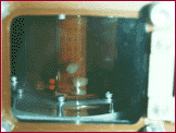
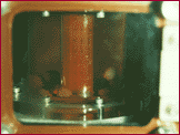
Flight and ground control studies (astronaut John Blaha, NASA-JSC team): Medium was recirculated between the bioreactor and the gas exchanger at 4 mL/min for 20 min four times per day, and 50 to 100 mL of fresh medium were infused into the system approximately once per day. As a result, medium metabolic parameters were maintained within previously established target ranges (i.e. pH between 6.9 and 7.4, partial pressure of oxygen between 71 and 127 mmHg, and glucose concentration between 3.0 and 4.3 g/L) in both groups for the duration of the study, as assessed using portable cartridges. Concentration gradients within the bioreactor were minimized by differential rotation of the inner and outer vessel walls at 10 and 1 rpm, respectively, in microgravity, and by the convection associated with gravitational construct settling during solid body rotation of the bioreactor at 28 rpm in unit gravity. In the Mir group, gas bubbles were observed in the bioreactor between flight days 40 and 130. The amount of gas stabilized at approximately 20% of the total bioreactor volume, and the bubbles did not appear to come into direct contact with the constructs, as assessed by videography. An equal amount of gas was introduced into the bioreactor in the Earth group, in order to match conditions on Mir as closely as possible.
Postflight (MIT): Constructs were assessed at the time of launch (i.e. after 3 months of culture) and after 4 additional months on either Mir or Earth (i.e. after 7 months of culture), and compared to full thickness natural calf articular cartilage. Constructs were assessed with respect to the following parameter: (1) size and morphology (weight, histological/ultrastructural appearance); (2) biochemical composition (DNA, glycosaminoglycan (GAG), collagen type II, total collagen); (3) viability of reisolated cells (trypan blue exclusion, intracellular esterase activity); (4) tissue metabolism (incorporation of radiolabeled tracers into macromolecular GAG and collagen); (5) mechanical properties in confined compression (aggregate modulus, dynamic stiffness, hydraulic permeability).
Results
Construct shape: Constructs grown on Mir tended to become more spherical while those grown on Earth maintained their initial discoid shape, as assessed histologically and from their respective aspect ratios (i.e. height to width) of 0.72±0.08 and 0.62±0.03 (p<0.05). These findings might be related to differences in cultivation conditions, i.e. videotapes showed that constructs floated freely in microgravity but settled and collided with the rotating vessel wall at 1-G.
Construct structure: Final samples from Mir and Earth appeared histologically cartilaginous throughout their entire cross-sections (5 - 8 mm thick), with the exception of fibrous outer capsules (0.15 - 0.45 mm thick), as assessed using safranin-0 stain for GAG and immunohistochemical staining for collagen type II. Constructs grown on Earth appeared to have a more organized extracellular matrix with more uniform collagen orientation as compared to constructs grown on Mir, but the average collagen fiber diameter was similar in the two groups (22±2 nm).
Construct composition: Constructs at the time of launch and after additional cultivation on Mir and on Earth contained 13±1, 14±0.8 and 19±0.2 million cells, respectively. On Earth, construct wet weights increased 1.7-fold between 3 and 7 months, which could be attributed to increasing amounts of cartilage-specific tissue components (i.e. GAG and collagen type II). In contrast, on Mir construct wet weights increased 1.3-fold over the same time interval, due to deposition of collagen and unspecified components that were not GAG. The polymer scaffold represented less than 0.3% of the final construct wet weight. The fraction of the total collagen that was type II decreased, but not significantly, from 92±19% at launch to 78±4% at landing, demonstrating relatively good maintenance of the chondrocytic phenotype.
Construct function: Construct mechanical properties improved both on Mir and on Earth resulting in an increase in aggregate modulus, HA, and a decrease in hydraulic permeability, k. Dynamic stiffness also increased with culture time and showed the characteristic frequency dependence of natural cartilage. Mechanical properties of Mir-grown constructs were inferior to those of Earth-grown constructs. Specifically, the aggregate modulus of Earth-grown constructs was indistinguishable from natural calf cartilage and was three-fold higher than that of Mir-grown constructs.
Tissue engineering is the creation of new tissues from component cells and biomaterial scaffolds. Our flight study was the first to demonstrate that engineered tissues can be grown for several months in space. Cartilaginous tissues grown on Mir were smaller, more spherical, and mechanically weaker than corresponding tissues grown on Earth. Our data is consistent with previous reports that space flight weakens the bones and muscles of experimental animals and humans. But, ours was the first controlled comparison of isolated tissues grown on space or Earth for period of several months. Further studies of tissue engineering in space might help us understand, prevent or treat conditions such as osteoporosis that affect astronauts as well as bedridden and elderly people on Earth.
Earth Benefits Publications
Freed, L.E., Pellis, N., Searby, N., de Luis, J., Preda, C., Bordonaro, J., Vunjak-Novakovic, G. Microgravity Cultivation of Cells and Tissues. Gravitational & Space Biology Bull. (invited article, submitted 10/98).
Principal Investigators Co-Investigators![]()
NASA-3
Preflight (MIT): Cell-polymer tissue "constructs" based on bovine calf articular chondrocytes and biodegradable polyglycolic acid scaffolds (5 million cells per 5 mm diameter x 2 mm thick scaffold) were cultured in rotating bioreactors for 3 months at 1-G prior to launch. Culture medium consisted of Dulbecco's modified Eagle medium with 4.5 g/L glucose, 10% fetal bovine serum, 10 mM HEPES buffer, 0.1 mM nonessential amino acids, 0.4 mM proline, 50 mg/L ascorbic acid, 50 U/mL penicillin, 50 mg/mL streptomycin, and 0.5 mg/mL fungizone. After 3 months, constructs were transferred into each of two flight-qualified rotating, perfused bioreactors (the Biotechnology system, BTS) for an additional 4 months of cultivation on either the Mir Space Station or on Earth. Specifically, one BTS containing ten constructs was transferred to Mir via the US Space Shuttle STS-79 (9/16/96 launch) and brought back to Earth via STS-81 (1/22/97 landing). A second BTS with ten constructs served as an otherwise identical study conducted on Earth, at JSC.
Cellular viability: Mir-grown constructs assessed 30 hours postflight were comparable to Earth-grown constructs with respect to cell viability and biosynthetic activity. Specifically, constructs from both groups consisted of 95-99% viable cells, as judged by trypan blue exclusion and by intracellular esterase activity, and incorporated radiolabeled tracers into macromolecules at comparable rates. The latter may represent a rebound in chondrocyte metabolism in the Mir-grown constructs, which were assayed following return from space.
The cartilage in space experiment improves our understanding of how gravity affects tissue development. Gravitational forces make it difficult to culture large aggregates of cells because the tissues sediment. This limits the direction of cellular growth to only two-dimensions. By studying tissues in microgravity, scientists can gain insight into tissue development as it occurs in the body and, they can grow tissues for longer periods of time. BTS cartilage provides a cellular model of the response to gravitational unloading. This can be used on the ground to better understand what happens to the musculoskeletal tissue response in immobilized patients, intrauterine development, and osteoporosis.
Tissue Engineering of Cartilage in Space. L.E. Freed, R. Langer, I. Martin, N.R. Pellis, G. Vunjak-Novakovic. Proc. Nat. Acad. Sci. U.S.A. 94: 13885-13890, 1997. This paper can be accessed at http://www.pnas.org. A commentary on this work appeared in the same PNAS issue on page 13380.
Lisa E. Freed, M.D., Ph.D.
Massachusetts Institute of Technology (MIT)
Gordana Vunjak-Novakovic, Ph.D.
Neal R. Pellis, Ph.D.
The NASA/Johnson Space Center team working on BTS
![]()
|
|
Curator:
Julie Oliveaux
Responsible NASA Official: John Uri |
Page last updated: 07/16/1999
.gif)